All information is based on current medical research (2020-2026). Written specifically for patient education by Dr. Antonio Gargiulo, reproductive medicine and advanced gynecologic surgery.
This is Part 1 of a two-part series on uterine fibroids. Part 2 — “Treating Uterine Fibroids: A Guide to Your Options” — covers all treatment choices in detail, including surgery, radiofrequency ablation, and uterine artery embolization.
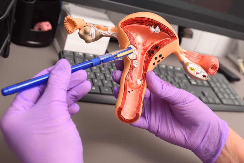
What Are Uterine Fibroids?
Uterine fibroids are non-cancerous growths that develop in or on the wall of the uterus (womb). They are made up of smooth muscle cells and fibrous connective tissue, and they are the most common benign tumors of the female reproductive system. Studies estimate that fibroids affect up to 70% of white women and more than 80% of women of African ancestry at some point during their lifetime, though many women never know they have them because they cause no symptoms at all.
Fibroids go by several names you may encounter in medical reports or conversations with your doctor: leiomyomas, myomas, or fibromyomas. They all mean the same thing. They are not cancer, they do not become cancer, and they do not spread to other organs.
Fibroids vary enormously in size, from tiny seedlings barely visible to the naked eye, to large masses that can distort the shape of the uterus and press on surrounding organs. They can grow as a single nodule or in clusters. Their location within or on the uterus matters greatly: fibroids that push into the inner cavity of the uterus tend to cause more bleeding and fertility problems, while those buried deep in the muscle wall or growing on the outer surface may cause pressure and bulk symptoms.
Could I Have Them? Recognizing the Symptoms
Fibroids are extremely common. One in two women over 40 have them. Hence they are considered a normal condition, not a disease, until they prove themselves otherwise. Between 50% and 75% of women with fibroids have no symptoms at all and discover them only during a routine ultrasound or pelvic exam. When symptoms do occur, they depend heavily on how large the fibroids are, how many there are, and exactly where they are located in the uterus.
Heavy menstrual bleeding is the most common complaint. Periods may be unusually long, very heavy, or filled with large clots. Over time, this blood loss can lead to iron-deficiency anemia — a shortage of iron in the blood that leaves you feeling exhausted, weak, dizzy, or short of breath even with ordinary activity.
Pelvic pain and pressure are also very common. You may feel a persistent dull ache, a sense of heaviness or fullness in the lower belly, or sharp cramping during your period. Some women describe it as a constant feeling of bloating that does not go away.
Painful intercourse can occur when fibroids press on or distort the structures involved in sexual activity.
Bladder and bowel symptoms happen when fibroids grow large enough to press on nearby organs. You may feel the need to urinate more often than usual, have trouble fully emptying your bladder, or experience constipation.
Lower back pain is reported by some women and can be mistaken for a muscle or spine problem.
Fertility and pregnancy problems — fibroids that distort the uterine cavity or block the entrance to the fallopian tubes can interfere with conception. Even after conception, certain fibroids have been linked to a higher risk of miscarriage, early delivery, or complications during pregnancy.
Spotting between periods — light bleeding outside of your regular menstrual cycle can also occur, particularly with fibroids that grow into or near the inner lining of the uterus.
One important thing to keep in mind: the size of a fibroid does not always predict how much trouble it causes. A small fibroid in the wrong location — pressing into the uterine cavity — can cause far more bleeding than a large one buried in the muscle wall. Your symptoms, not the size alone, are what guide the need for treatment.
What Causes Fibroids?
The exact cause of uterine fibroids is not fully understood, but research has identified several biological and environmental factors that drive their development and growth.
Hormonal influence. Fibroids are strongly fueled by the female hormones estrogen and progesterone. They grow during the reproductive years when these hormones are active, they often enlarge during pregnancy when hormone levels are high, and they typically shrink after menopause when hormone production falls. This hormonal dependence is why most medical treatments for fibroids work by reducing hormone levels.
Genetic factors. Fibroids develop from a single abnormal uterine muscle cell that multiplies. Specific gene mutations — particularly in genes called MED12 and HMGA2 — have been found in a large proportion of fibroids and are thought to play a central role in their formation. A family history of fibroids significantly increases your own risk.
Inflammation and other molecular pathways. Beyond hormones, chronic low-grade inflammation within the uterus, along with various growth-promoting proteins and molecular signaling pathways, appears to contribute to fibroid development and growth. Researchers are actively studying these non-hormonal mechanisms as potential targets for new treatments.
Known risk factors include being of reproductive age (particularly between 30 and 50 years), a family history of fibroids, being of African descent (fibroids are more common, appear earlier, and tend to be more severe in Black women), obesity, early onset of menstrual periods, a diet high in red meat and low in green vegetables and fruit, possible exposure to environmental chemicals that interfere with hormone signaling, and a history of prior uterine surgery.
How Are Fibroids Diagnosed?
1. Medical history and physical exam. Your doctor will begin by asking about your periods, any pain or pressure symptoms, your reproductive history, and whether fibroids run in your family. During a pelvic exam, an enlarged or irregularly shaped uterus may suggest fibroids — but imaging is needed to confirm the diagnosis.
2. Transvaginal ultrasound — the first-line screening test. A small, smooth probe placed gently inside the vagina produces detailed sound-wave images of the uterus. This is safe, widely available, affordable, and involves no radiation. It is the recommended starting point for fibroid evaluation. A 2024 review published in *Fertility and Sterility* confirmed that transvaginal ultrasound remains the recommended first-line diagnostic tool, though its accuracy depends heavily on the operator and the complexity of the case.
3. MRI – A FUNDAMENTAL PREOPERATIVE TEST. MRI uses magnetic fields — no radiation — to produce highly detailed images of the uterus and its contents. In a 2023 invited commentary published in RadioGraphics, Dr. Fiona Fennessy (Department of Radiology, Harvard Medical School) and Dr. Antonio Gargiulo (Department of Obstetrics and Gynecology, Harvard Medical School) argued that pelvic MRI has now become the basis of personalized fibroid care. Their core message: fibroid management cannot be guided by a one-size-fits-all approach. Each patient’s fibroid burden is unique in location, size, number, and relationship to the uterine cavity, and treatment decisions must be grounded in precise, high-quality imaging. MRI also enables careful distinction between fibroid subtypes, between fibroids and adenomyosis, and — critically — between benign fibroids and the rare but serious uterine sarcoma that can mimic them on imaging.
When is A Surgical Procedure Used for Diagnosis?
In some cases, a hysteroscopy — a thin camera inserted through the vagina and cervix into the uterine cavity — may be used to directly visualize and in some cases simultaneously remove fibroids growing inside the cavity. Laparoscopy (a camera inserted through the belly button) may occasionally be used as part of surgical planning, but it is not a primary diagnostic tool for fibroids.
Fibroids versus other conditions. Fibroids share many symptoms with adenomyosis and endometriosis, and these conditions frequently coexist in the same patient. Accurate imaging by an experienced specialist is essential to distinguish between them, because they respond to different treatments.
The Decision to Watch and Wait — and When Waiting Becomes Dangerous
If you have fibroids but no symptoms, or only mild symptoms that do not affect your quality of life, watchful waiting with regular monitoring is a reasonable approach for many women — particularly for those who have already completed childbearing or are approaching menopause, where natural hormonal changes will cause fibroids to shrink on their own.
But watchful waiting requires more than just waiting. As Dr. Fiona Fennessy and Dr. Antonio Gargiulo argued in their 2023 invited commentary in RadioGraphics, pelvic MRI has now become the basis of personalized fibroid care. The decision to observe rather than treat must itself be grounded in precise imaging — knowing exactly what you have, where it is, and tracking whether it changes over time.
For a woman who has not completed her childbearing, this point carries urgent weight. Watching a fibroid grow year after year without systematic imaging surveillance is not watchful waiting — it is uninformed delay. Fibroids grow predictably under the influence of estrogen, and their growth is not always gradual. Volume can expand rapidly and unpredictably. What begins as a small fibroid unlikely to affect reproduction can become a significantly larger tumor that compromises the structural integrity of the uterine wall itself — reducing muscle reserve, increasing surgical complexity, and in the worst cases, making a safe uterus-preserving operation impossible. Countless uteri have been lost because watchful surveillance became complacent, missing a period of rapid tumor growth until surgery was no longer feasible without removing the uterus entirely. If you have fibroids and you have not yet had all the children you want, the decision to “watch and wait” must be made actively, carefully, with regular MRI-based re-evaluation — and never allowed to drift into indefinite delay.
Frequently Asked Questions
1. Do fibroids go away on their own?
Fibroids do not usually disappear on their own, but they typically shrink after menopause when estrogen levels fall. Small, asymptomatic fibroids may remain stable for years without causing any problems.
2. Is this the same as endometriosis?
No. Fibroids are non-cancerous muscle growths within or on the uterus. Endometriosis is a separate condition in which tissue similar to the uterine lining grows outside the uterus. These conditions can coexist, and their symptoms overlap, which is why a careful evaluation is important.
3. Can fibroids affect my fertility?
Yes, in some cases — particularly fibroids that grow into or distort the uterine cavity. Many women with fibroids conceive without difficulty, but the location of the fibroid matters more than its size.
4. What imaging should I ask my doctor about?
A transvaginal ultrasound is the right first step. If results are unclear, if multiple or complex fibroids are suspected, or if surgery is being planned, an MRI will give your doctor the most accurate and complete picture — and as Dr. Fennessy and Dr. Gargiulo have argued, it is now the foundation of truly personalized fibroid care.
Ready to learn about your treatment options? Continue to Part 2: “Treating Uterine Fibroids: A Guide to Your Options” — covering medications, myomectomy, radiofrequency ablation (the Sonata procedure), and uterine artery embolization, with a frank discussion of which options are and are not appropriate for women who have not completed childbearing.*
Sources We Used
So You Can Read Them, Question Them, and Decide for Yourself
We believe that informed patients are empowered patients. In an age where artificial intelligence and open-access science place original research within reach of anyone, you have every right to go to the source, read it yourself, and form your own conclusions. Patient education on this website is taken seriously: we do not simplify at the cost of truth, and we do not ask you to take our word for it.
Every statement in this article carries two layers of accountability. It has been filtered through the critical eye of Dr. Antonio Gargiulo, drawing on four decades of clinical and surgical experience in reproductive medicine and advanced gynecologic surgery. And it is independently traceable to a peer-reviewed scientific publication, listed below with its full reference and digital identifier (DOI), so you can retrieve and read the original source at any time.
We see healthcare as a shared responsibility between doctors and patients. Shared responsibility requires shared access to information. These references are not a formality. They are here for you.
1. Giuliani E, As-Sanie S, Marsh E. Epidemiology and management of uterine fibroids. International Journal of Gynaecology and Obstetrics. 2020. DOI: 10.1002/ijgo.13102
2. Munro MG, Tchaikovski S, Murji A. The epidemiology and pathogenesis of uterine fibroids.International Journal of Gynaecology and Obstetrics. 2025. DOI: 10.1002/ijgo.70527
3. Dolmans M, Petraglia F, Catherino W, Donnez J. Pathogenesis of uterine fibroids: current understanding and future directions. Fertility and Sterility. 2024. DOI: 10.1016/j.fertnstert.2024.02.048
4. Mension E, Carmona F, Vannuccini S, Chapron C. Clinical signs and diagnosis of fibroids from adolescence to menopause. Fertility and Sterility. 2024. DOI: 10.1016/j.fertnstert.2024.05.003
5. Lakabi R, Harth S, Meinhold-Heerlein I, et al. Diagnosis and classification of uterine fibroids. International Journal of Gynaecology and Obstetrics. 2025. DOI: 10.1002/ijgo.70538
6. Fennessy FM, Gargiulo AR. Invited Commentary: Pelvic MRI Is Now the Basis of Personalized Fibroid Care. RadioGraphics. 2023;43(6). DOI: 10.1148/rg.220212
7. Ashraf S, Asim A, Taj A, et al. Diagnostic Accuracy of Pelvic MRI and Transvaginal Ultrasound for Detecting Uterine Fibroids and Adenomyosis. Pakistan Journal of Medical and Health Sciences. 2024. DOI: 10.53350/pjmhs02024181462
8. Ascher S, Wasnik A, Robbins J, et al. ACR Appropriateness Criteria: Fibroids. Journal of the American College of Radiology. 2022. DOI: 10.1016/j.jacr.2022.09.019
9. Kayastha B, Shrestha B, Pradhan A. Clinical Presentation of Uterine Fibroid and the Presence of Co-Existing Other Pathologies Like Endometriosis and Adenomyosis at Tertiary Care Centre. Journal of College of Medical Sciences — Nepal. 2025. DOI: 10.3126/jcmsn.v21i4.86260
This article is intended for general patient education. It does not replace a consultation with your gynecologist or healthcare provider.