


All information is based on current medical research (2018–2026). Written specifically for patient education by Dr. Antonio Gargiulo (reproductive surgeon and endometriosis excision specialist) and Dr. Ammara Watkins (thoracic surgeon, Lahey Hospital & Medical Center). This article does not replace a consultation with your gynecologist, endometriosis surgeon, or thoracic surgeon.
A Spot on the Diaphragm — Now What?
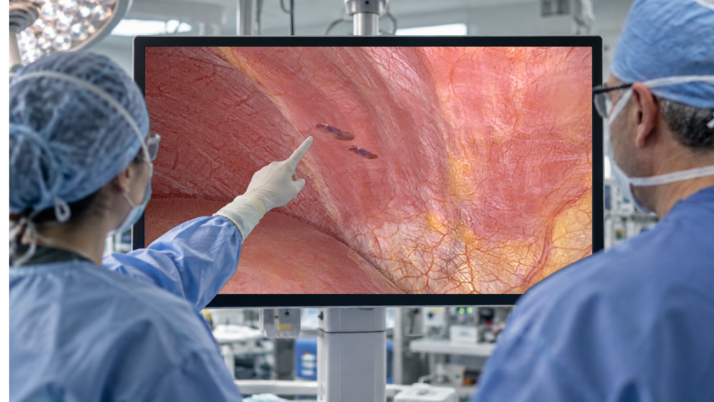
You’re on the table for pelvic endometriosis surgery. It’s going well. Then, at the very top of the abdomen, the surgeon looks up at the underside of the diaphragm — the dome-shaped breathing muscle between your belly and your chest — and sees a few small brown or blue spots. Endometriosis. You’ve never had chest pain, shoulder pain, or trouble breathing. The spots aren’t bothering you at all.
Remove them while you’re already undergoing surgery? Or leave them exactly where they are?
This scenario is not rare; it is hard to predict even with the best radiologic screening, and is one of the genuinely unsettled decisions in endometriosis surgery: that is why we wrote this together. One of us (Dr. Gargiulo) looks up at the diaphragm from below, during pelvic surgery. The other (Dr. Watkins) is a thoracic surgeon who works the other side of that same muscle, looking down from inside the chest. We see the same disease from opposite directions and don’t always agree on what to do about it.
The one idea to carry through: a diaphragmatic lesion that causes no symptoms rarely progresses and can usually be left alone — and no evidence shows that removing it helps. The evidence is thin, and the right call depends on where the lesion sits and how much surgery removing it would take.
What Diaphragmatic Endometriosis Actually Is
Endometriosis is tissue resembling the uterine lining growing where it doesn’t belong. Usually it stays in the pelvis. In about 1 in 10 patients, it turns up outside the pelvis, and after the abdominal wall, the diaphragm is one of the most common of those sites[1]. It’s uncommon: across surgical series it shows up in roughly 0.7% to 4.7% of endometriosis patients[1, 2], and in 1.86% of women in one large single-center series[2].
Three features define it, and each one drives the decision ahead:
- It’s overwhelmingly right-sided. Across the published cases, diaphragmatic lesions sit on the right in about two-thirds of women, and when only one side is involved, 90% land on the right[3]. The reason is plumbing: abdominal fluid circulates in a clockwise current that sweeps cells up the right side, where the liver’s anchoring ligament (the falciform ligament) acts like a fence and holds them there[1].
- Most lesions are shallow. The majority sit on the surface lining; nodules that burrow into the muscle are less common[1]. In one series, only 27% had deep infiltration, and full-thickness disease — where the surgeon has to open all the way through the muscle — was present in under 17%[2].
- It travels with worse pelvic disease. Diaphragmatic endometriosis rarely occurs alone, and its presence usually signals advanced, deeply infiltrating disease elsewhere[1]. In that single-center series, 87% of these women had deep infiltrating pelvic endometriosis and about half had adenomyosis[2]. Roughly half also had infertility[1, 2].
What this means for you: A diaphragmatic lesion is partly a message. Even a silent spot tells your team to look hard at the pelvis, because the disease that matters is usually down there.
Why These Lesions Are Usually Silent
Most diaphragmatic endometriosis causes no symptoms at all[1]. It’s usually found by accident — a surprise spotted during a laparoscopy done for pelvic disease[2].
This article is about exactly that patient: a woman with a silent, incidentally discovered lesion and no chest, shoulder, or upper-abdominal complaints. When a diaphragmatic lesion does cause symptoms, that’s a different clinical problem with a different answer — the subject of the companion article flagged at the end.
What this means for you: An incidentally found lesion with no symptoms is behaving the way most of them do. That silence is information, not a problem to reflexively fix.
The Central Question — and What the Evidence Says
When a lesion causes no symptoms, two claims get made, and they are not the same:
- “Asymptomatic lesions rarely progress, so we can safely leave them.”
- “Operating on asymptomatic lesions prevents future problems, so we should remove them.”
The literature backs the first and not the second.
Reviews conclude that asymptomatic diaphragmatic endometriosis is very unlikely to progress and can be left in place when found incidentally[1]. In the single-center series, all four patients whose lesions were left alone stayed stable[2]. Endometriosis generally behaves the same way: followed without treatment, most lesions stay stable or shrink rather than grow. A 2024 systematic review and meta-analysis found untreated deep endometriosis progressed in only about one in five women (21.4%)[4], and a 2023 expectant-management cohort of 135 women found the disease static in half and regressing in another 13%, leaving only about a third that grew at all[5]. These are studies of pelvic disease, so extending them to the diaphragm is a reasonable extrapolation, not proof.
The second claim has no real support. Removing a silent diaphragmatic lesion has never been shown to prevent future problems or improve any long-term outcome. Those studies simply haven’t been done.
Better support is not proven. The published experience of deliberately watching asymptomatic diaphragmatic disease is a handful of patients — four stable in one series, two of three in another.[1, 2] Follow-up ran only 6 to 14 months, too short to rule out slow progression[2]. When a surgeon tells you untreated patients “remained stable,” that’s how few patients and how few months the reassurance rests on.
The guidelines agree. The 2022 ESHRE endometriosis guideline states that clinicians should not routinely remove or ablate an incidental finding of asymptomatic endometriosis at the time of surgery[6].
FROM THE TWO SIDES OF THE DIAPHRAGM
Gargiulo — View from Below: When I glance up at a quiet spot on the right diaphragm during pelvic surgery, my instinct is restraint. It isn’t hurting her; it probably won’t, and the guideline tells me not to chase it. That day, my job is the pelvis.
Watkins — View from Above: My caution is the mirror image. From the chest side, a spot that looks small and superficial from below can hide a full-thickness hole, or a lesion on the pleura you can’t see from the abdomen. I’m not arguing to remove silent disease. I’m arguing that “we looked and it was small” from one side of the muscle isn’t the whole picture. Additionally, I have found that thoracic endometriosis is not always asymptomatic. Instead, patients may become accustomed to or normalize symptoms associated with the disease, often failing to recognize their significance until specifically asked.
What this means for you: For a silent lesion, “leave it alone” is the evidence-based default. If a surgeon wants to remove one anyway, have that conversation before surgery — and know it’s a judgment call, not a proven benefit.
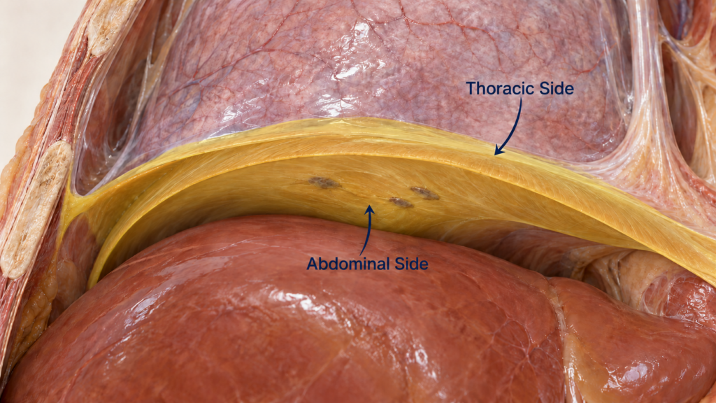
Figure 1. The diaphragm viewed from two sides. This cutaway shows the right lung above the diaphragm and the liver below it. The diaphragm is artificially highlighted in yellow for ease of identification by the reader. Three small endometriotic implants are illustrated on its abdominal surface, where they may be seen during pelvic surgery. The arrows show the opposing abdominal and thoracic viewpoints: a clear view from one side does not necessarily reveal what is present on the other. Anatomical illustration; not an operative photograph.
The Seven Faces of a Diaphragm Lesion
“Diaphragmatic endometriosis” is one label for situations that run from trivial to formidable, and the decision changes with each:
- Small superficial implants. Shallow surface spots — the most common, and the easiest to treat lightly or leave.
- Larger or multifocal disease. More lesions, more surface, harder to clear.
- Deep or full-thickness disease. The lesion goes through the muscle. Removing it means entering the chest and repairing the hole.
- Posterior right-sided disease hidden behind the liver. The most treacherous spot. Much of the right diaphragm hides behind the liver, so lesions there are invisible unless the surgeon mobilizes the liver to look[1]. A spot on the front can be a “sentinel” pointing to worse disease behind[1].
- Disease that reaches the chest side. Lesions extending onto the lung lining (pleura) or perforating the diaphragm — anatomically the most extensive form, and the one a gynecologic surgeon cannot fully assess or reach from the abdomen alone.
- Incidental disease found during pelvic surgery. The classic silent-spot dilemma.
- Disease known before surgery from imaging. MRI occasionally flags it in advance, but it’s imperfect here: sensitivity around 78–83% in the best study, below 50% in others, and it underestimates how far the disease extends[1]. For a broader discussion of diagnostic methods and their limitations, read our article on new tests for diagnosing endometriosis without surgery.
What this means for you: Ask your surgeon which of these you have. A superficial anterior spot and a full-thickness lesion behind the liver share a name and nothing else, and they deserve different decisions.
Why Treat Disease That Doesn’t Hurt? — and Why Not?
Why treat it
- You’re already in there. If the belly is accessible and the lesion is superficial and easy to reach, handling it now might spare a second operation.
- A tissue diagnosis. Removing it confirms what it is.
- Possible prevention. Clearing disease early might stop it becoming the harder, full-thickness problem — biologically plausible, but unproven.
- Completeness. Incomplete endometriosis surgery is linked to higher recurrence, so some surgeons clear everything they see[1].
Why not treat it
The diaphragm is a dangerous place to operate:
- A collapsed lung (pneumothorax). Cutting through or near the diaphragm can let air into the chest.
- Liver injury. Reaching the back of the right diaphragm means mobilizing the liver. In one robotic series, this caused hemorrhage severe enough to convert to open surgery[1].
- Holes and hernias. Full-thickness removal leaves a defect to repair; long-term complications in one series included three cases of the liver herniating up through the diaphragm and a pneumothorax[1].
- Phrenic nerve damage. The nerve driving the diaphragm runs across it; injuring it can weaken breathing permanently.
- Causing the symptoms you meant to prevent. Surgery near the diaphragm can create new chronic pain from nerve irritation or scarring. In the single-center series, two patients developed new shoulder pain afterward[2].
Done well, the diaphragm-specific complication rate is low, and reoperation is uncommon — about 4% in one series[2], with a 1.7% intraoperative and 6.6% long-term complication rate in the largest robotic series[1]. The surgery isn’t reckless. But every one of these risks, however small, is being run against a lesion that is doing nothing — a hard trade to justify.
And the reason not to rush is that the disease, left alone, mostly stays put. In the expectant-management data, half of untreated deep-endometriosis lesions stayed exactly the same and another 13% shrank; only about a third grew at all[5].
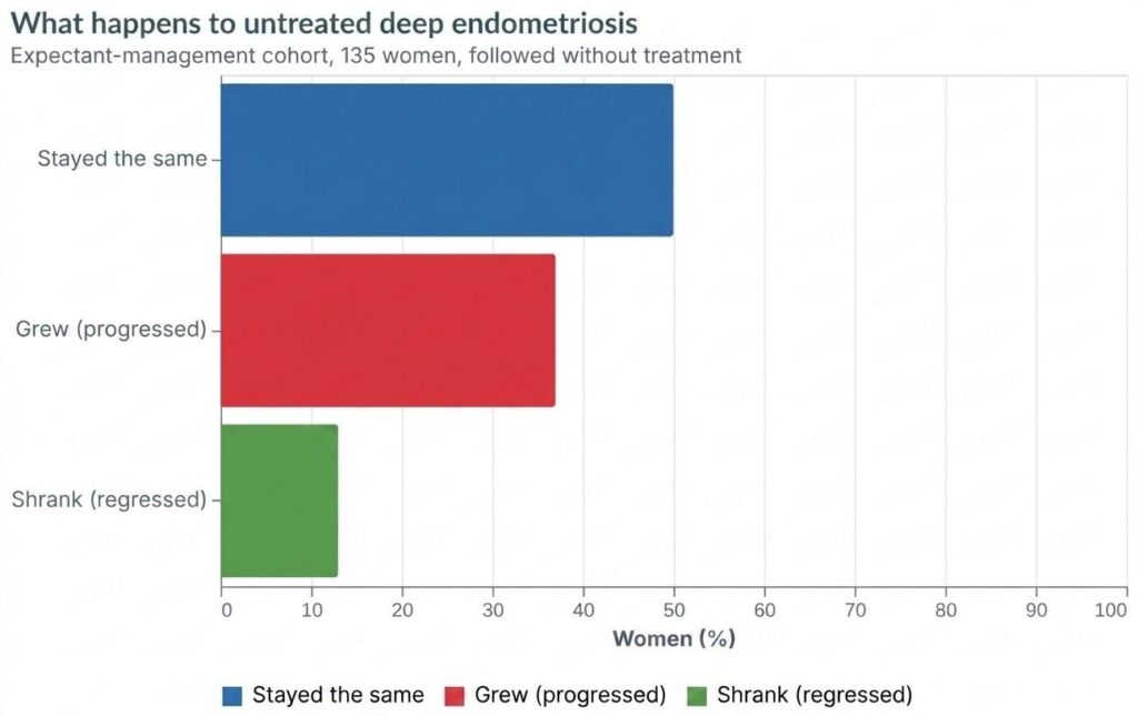
Figure 2. In a 2023 expectant-management cohort of 135 women with deep pelvic endometriosis followed without treatment, half the lesions stayed the same and 13% shrank, while about a third grew.
FROM THE TWO SIDES OF THE DIAPHRAGM
Watkins — View from Above: As a thoracic surgeon, I am comfortable managing the technical challenges of these procedures, whether that involves entering the pleural space, mobilizing the liver, or repairing a diaphragmatic defect. What gives me greater pause is the possibility of introducing a complication that could have lasting consequences. Even when we can safely perform an intervention, we need to be certain that the anticipated benefit justifies the risks.
Gargiulo — View from Below: Which is why my threshold to touch the diaphragm rises the moment a lesion is deep, posterior, or behind the liver. A superficial anterior spot I can ablate in thirty seconds. A full-thickness lesion that means opening the chest is a different operation — and in a woman with no symptoms, the math almost never favors it.
What this means for you: The question isn’t “is there disease?” It’s “what will removing it cost, and what do I get back?” For silent disease, usually: too much cost, too little return.
What About Fertility and IVF?
About half of women with diaphragmatic endometriosis are infertile — 52% in the largest single-center series[2], and the review literature puts it at roughly half across cohorts[1]. They carry higher infertility rates than endometriosis patients without diaphragmatic involvement[2].
The diaphragm isn’t the cause, though. These lesions almost never occur alone — they mark advanced, deeply infiltrating pelvic disease, and it’s that pelvic burden (endometriomas, deep nodules, adhesions) that lowers fertility. Diaphragmatic disease, deep pelvic endometriosis, and infertility travel together as one severe subtype[1, 2].
For patients with ovarian endometriomas who are considering fertility preservation, our guide to egg retrieval before endometrioma surgery provides additional information to discuss with their fertility specialist.
After surgery, the fertility numbers are thin but not discouraging. In that single-center series, 9 of 11 infertile women tried to conceive and 4 succeeded — a 44% clinical pregnancy rate, three spontaneously and one by ICSI[2]. But those women had their pelvic disease resected at the same operation, so the result can’t be credited to the diaphragmatic part. No study shows that removing a diaphragmatic lesion on its own improves fertility, and it’s unclear whether treating superficial lesions helps pain or fertility at all[1].
What this means for you: If you have a silent diaphragmatic lesion and want to conceive, the fertility plan should target your pelvic disease and the usual paths — timed conception, pelvic surgery, or IVF/ICSI — not the diaphragmatic spot. Removing it won’t improve your odds of pregnancy, so it shouldn’t be done for fertility’s sake.
A Practical Way to Decide
There’s no clean “always remove / never remove” rule for a silent lesion. What we can give you is a principle and the factors that move it.
The principle: the threshold to remove asymptomatic disease should rise with the amount of surgery it takes to remove it. A superficial spot you could safely ablate in seconds is one thing; a full-thickness lesion behind the liver that means opening the chest and mobilizing the liver is another. The bigger the operation, the stronger the case for leaving a silent lesion exactly where it is. Treat this as a working framework, not a guideline.
Stronger case to leave it alone: deep, posterior, or behind-the-liver location; disease that would need the chest opened or the liver mobilized to reach; any doubt about whether it could be cleared safely; a preference to avoid added surgical risk.
The only reasons to consider removing a silent lesion: it’s superficial and genuinely trivial to deal with while you’re already operating in the pelvis; you specifically want a tissue diagnosis; or you’ve thought it through in advance and chosen to. None of these is a proven benefit — each is a judgment call to make before surgery, not on the spot.
The one rule we both hold: a silent, incidental lesion should be left alone, and a planned pelvic operation should not turn into diaphragmatic surgery on the spot. That is what the evidence and the 2022 ESHRE guideline both say[6].
What this means for you: Ask your surgeon, before the operation, what they will do if they find a silent diaphragmatic spot. The good decisions get made with you, in advance — not improvised while you’re asleep.
Our Take: Two Surgeons, One Answer
We came at this from opposite sides of the same muscle and land in the same place, because the evidence points one way. For a silent, incidentally found lesion, restraint wins. It probably won’t progress[5], removing it has never been shown to help, and the diaphragm punishes unnecessary heroics. Leaving it alone is the evidence-based, guideline-concordant default[6] — and a surgeon who removes one anyway should be able to explain why in your specific case and call it what it is: a judgment call, not a proven benefit.
Where we lean differently is worth hearing. Dr. Gargiulo’s instinct is to protect the diaphragm and reserve any operation on it for a clear reason. Dr. Watkins’s instinct is to make sure nobody calls a lesion “small and superficial” from below without appreciating what can hide on the chest side. Both instincts are protective; they simply guard against different mistakes. And we’re honest about the biggest gap: the natural history of untreated asymptomatic diaphragmatic disease rests on a handful of patients followed for months, not years[1, 2]. “Probably won’t progress” is the best-supported answer we have — not a guarantee.
A Note on Not Being Rushed
The most important thing about a silent diaphragmatic lesion is that it gives you time. Nothing about it demands a decision in the moment. If your surgeon finds one and raises the question of removing it, that is a conversation to have with you — awake, with the trade-offs laid out — not a call to make while you’re under anesthesia. Leaving a quiet lesion alone is not neglect; it is what the evidence and the guidelines support, and it keeps every option open.
Sources We Used
So you can read them, question them, and decide for yourself. We drew on the state-of-the-art review of diaphragmatic endometriosis, the largest single-center surgical series, a meta-analysis of lesion distribution, two studies of the natural history of untreated endometriosis, and the 2022 ESHRE guideline — and we’ve been explicit about where the evidence is thin, especially the natural history of untreated asymptomatic disease.
- Naem A, Roman H, Martin DC, Krentel H. A bird-eye view of diaphragmatic endometriosis: current practices and future perspectives. Frontiers in Medicine. 2024. DOI: 10.3389/fmed.2024.1505399
- Naem A, Andrikos A, Constantin A, et al. Diaphragmatic Endometriosis—A Single-Center Retrospective Analysis of the Patients’ Demographics, Symptomatology, and Long-Term Treatment Outcomes. Journal of Clinical Medicine. 2023. DOI: 10.3390/jcm12206455
- Vercellini P, Abbiati A, Viganò P, et al. Asymmetry in distribution of diaphragmatic endometriotic lesions: evidence in favour of the menstrual reflux theory. Human Reproduction. 2007. DOI: 10.1093/humrep/dem224
- Bandini V, Giola F, Ambruoso D, Cipriani S, Chiaffarino F, Vercellini P. The natural evolution of untreated deep endometriosis and the effect of hormonal suppression: a systematic literature review and meta-analysis. Acta Obstetricia et Gynecologica Scandinavica. 2024. DOI: 10.1111/aogs.14887
- Knez J, Bean E, Nijjar S, Tellum T, Chaggar P, Jurković D. Natural progression of deep pelvic endometriosis in women who opt for expectant management. Acta Obstetricia et Gynecologica Scandinavica. 2023. DOI: 10.1111/aogs.14491
- Becker CM, Bokor A, Heikinheimo O, et al. ESHRE guideline: endometriosis. Human Reproduction Open. 2022. DOI: 10.1093/hropen/hoac009
A note on terms and availability in the United States: Two words used above are worth defining. Ablation means destroying a lesion in place with heat or energy; excision means cutting it out. Both are standard, widely available laparoscopic techniques in the U.S. There is no drug or device FDA-approved specifically for diaphragmatic endometriosis — management is either surgical or, for a silent lesion, watchful observation, an approach reflected in international guidelines rather than in any product label.
References
1. Naem A, Roman H, Martin DC, Krentel H (2024) A bird-eye view of diaphragmatic endometriosis: current practices and future perspectives. Frontiers in Medicine. https://doi.org/10.3389/fmed.2024.1505399
2. Naem A, Andrikos A, Constantin A, et al (2023) Diaphragmatic Endometriosis—A Single-Center Retrospective Analysis of the Patients’ Demographics, Symptomatology, and Long-Term Treatment Outcomes. Journal of Clinical Medicine. https://doi.org/10.3390/jcm12206455
3. Vercellini P Asymmetry in distribution of diaphragmatic endometriotic lesions: evidence in favour of the menstrual reflux theory. https://air.unimi.it/handle/2434/34318
4. Bandini V, Giola F, Ambruoso D, et al (2024) The natural evolution of untreated deep endometriosis and the effect of hormonal suppression: A systematic literature review and meta‐analysis. Acta Obstetricia et Gynecologica Scandinavica. https://doi.org/10.1111/aogs.14887
5. Knez J, Bean E, Nijjar S, et al (2023) Natural progression of deep pelvic endometriosis in women who opt for expectant management. Acta Obstetricia et Gynecologica Scandinavica. https://doi.org/10.1111/aogs.14491
6. Vermeulen N Endometriosis. https://www.eshre.eu/-/media/sitecore-files/Guidelines/Endometriosis/ESHRE-GUIDELINE-ENDOMETRIOSIS-2022_2.pdf?hash=E1628E24D374F3EE6C9651EDB33235F8EB8ACBDA&la=en